
Estimated reading time: 7 minutes
Clubfoot, also called Congenital Talipes Equinovarus (CTEV), is considered one of the most common inherited lower limb deformities, which is generally seen in newborns. It is a structural deformity that was present at the time of birth, but if it is not recognized and treated appropriately, it can lead to lifelong disability. For taking concern for this condition clinically, everyone should understand its anatomical components, along with history-taking techniques, examination protocols, and evaluation protocols in detail.
What Is Clubfoot?
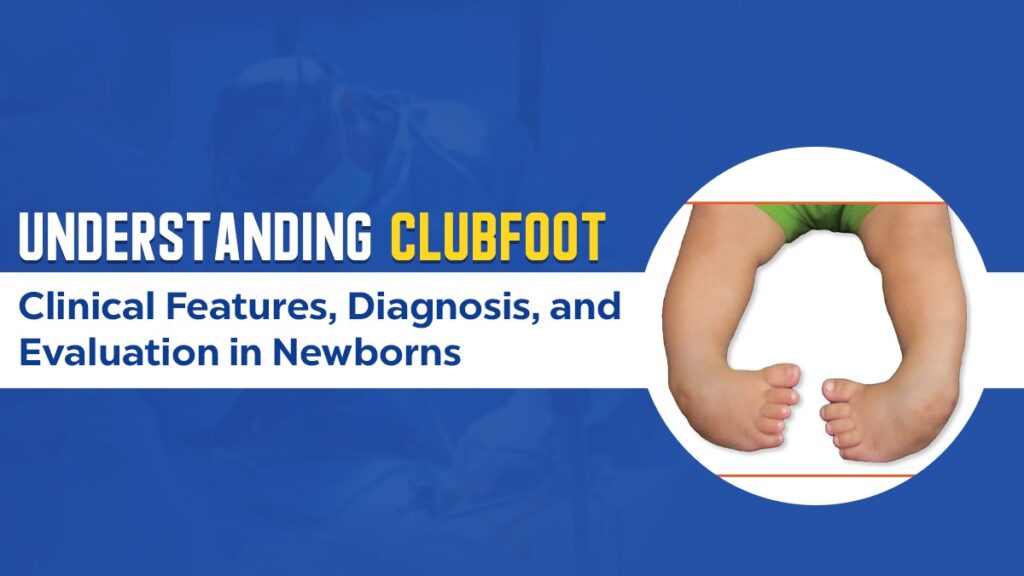
The Clubfoot is basically called a congenital structural deformity of the foot, which is distinguished by a combination of four distinct components that are:
- Cavus – it is called a high medial longitudinal arch, primarily a deformity of the midfoot.
- Adductus – the adductus is basically the Medial deviation of the forefoot.
- Varus – this means the Inversion or inward turning of the hindfoot.
- Equinus – the Plantar has been a flexion for deformity at the ankle joint, where the heel does not touch the ground.
Although these deformities now provide the classic appearance of a “twisted-in” foot. This helps to understand which segment is basically involved either it’s (midfoot, forefoot, hindfoot, or ankle) helps guide correction and prognosis.
Detailed History Taking
There are three forms of the foundation of diagnosis called antenatal, perinatal, and postnatal history.
1. Antenatal History
- Family History: Important to understand whether there’s any hereditary pattern or not, as clubfoot nowadays runs in family members due to some autosomal dominant tendencies.
- Oligohydramnios: This means the reduction of amniotic fluid, which can restrict fetal movement and easily lead to positional deformities.
- Decreased Fetal Movements: the reduction of fetal movement of suggestive of possible neuromuscular disorders.
- Congenital Anomalies on Scan: You know that antenatal scans play a very crucial role in ruling out syndromic or conditions associated with some anomalies like myelomeningocele or arthrogryposis.
2. Perinatal History
- Mode of Delivery: Behind presentation or any difficult labor can sometimes be connected with musculoskeletal malformation.
- Birth Weight and Neonatal Distress: When there is a problem of low birth weight or NICU admission, it may be associated with some systemic issues.
- Differential Diagnoses: There are some conditions, like cerebral palsy, which mimic talipes, the deformity that takes after the clubfoot.
3. Postnatal History
- The screening, which happens for spinal or limb anomalies, such as spina bifida, myelomeningocele, or hip dislocation.
- Checking for bladder atrophy or a tuft of hair on the lower back, which may point toward underlying neural tube defects.
Developmental and Family Factors
Although developmental milestones are not immediately relevant in newborns, assessing muscle tone and symmetry helps rule out neurogenic causes.
- Arthrogryposis and spina bifida may present with altered tone and rigid deformities.
- In the case of twin infants and facial expressions dysmorphism with “whistling face,” this may indicate the proper syndromic clubfoot.
These alliances typically remind all clinicians to appraise beyond the foot and consider the systemic and neurological involvement.
General Physical Examination
A meticulous general examination provides crucial diagnostic clues.
- Limb Symmetry: It basically determines whether the deformity is unilateral or bilateral in the child’s body.
- Facial Features: when we talk about the facial expressions, while look for syndromic features such as a flattened nasal bridge or a whistling face.
- Spine and Skin:
- Neurocutaneous markers: a clump of hair, a blessed dimple, or open spinal defects that perfectly suggest spina bifida or myelomeningocele.
- Bladder exstrophy, this one and other anterior abdominal wall defects, should also be ruled out.
Local Examination of the Foot
1. Inspection
- Laterality: Unilateral or bilateral involvement.
- Shape of the Foot:
- Medial crease: Deep and visible.
- Posterior crease: Extends laterally, suggesting severe deformity.
- Heel Pad: May appear small, displaced, or absent.
- Forefoot: Often adducted and pronated (needs supination during correction).
- Midfoot: Exhibits cavus deformity.
- Ankle: In equinus, the heel does not touch the surface.
- Skin: Initially normal, but may develop blisters or raw areas during plaster correction due to pressure points (especially near the talar head).
2. Palpation
- Muscle Bulk: Assess for calf muscle atrophy, a common finding, especially in unilateral cases.
- Calf Girth: Usually smaller on the affected side.
- Tendo-Achilles Tightness: It basically indicates the degree of ankle equinus deformity.
- Talus Head: In this, the Prominent dorsally in severe deformities.
- Navicular Bone: Medially displaced and palpable in rigid cases.
3. Assessment of Flexibility
The utmost flexibility helps to determine that the deformity is postural, which is also fully correctable. or structural (fixed).
- Forefoot Correction: first to check that the adduction is passively correctable or not.
- Heel Varus Correction: After that need to assess if the hindfoot can return to neutral.
- Ankle Movement: In the ankle movement it is assess the flexibility of equinus is assessed.
You know by the gentle manipulation that the positional clubfoot will be fully correctable by it, also it allows the foot to ankle flexion until the dorsum touches the shin, and it indicates a favorable prognosis.
Differentiating True Clubfoot from Other Conditions
It is essential to rule out neuromuscular and syndromic causes, such as:
- Arthrogryposis: Characterized by multiple joint contractures and stiff deformities.
- Spina Bifida and Myelomeningocele: Often associated with rigid, resistant deformities and neurological deficits.
These conditions usually require prolonged therapy, intensive physiotherapy, and sometimes surgical intervention for satisfactory correction.
Documentation and Scoring
The Baseline quantification is really critical for monitoring progress during treatment:
- The main and major thing is that the foot length and calf circumference should be recorded.
- The photographs, which are at the initial stage and follow-up stages are provide a visual record.
- Skin creases, toe alignment, and heel position, these three things are highly documented.
The Pirani Scoring System. This is basically very widely used for this purpose. It helps to assess six clinical signs, which are placed across three areas of the foot, midfoot, and hindfoot, as it really helps to quantify the danger and monitor the improvement after each stage of manipulation or casting.
Role of Radiological Evaluation
In neonates, radiographs are not recommended as the bones are largely unossified, making X-rays unreliable.
- Radiographs become relevant after six months of age, especially in:
- Uncorrected or relapsed clubfoot.
- Atypical or syndromic cases.
- Important radiological angles such as talocalcaneal, Meary’s, and kite’s angle are useful only in older infants or during surgical planning.
Summary
| Aspect | Key Points |
| Definition | Congenital structural deformity with cavus, adductus, varus, and equinus components |
| History | Antenatal (family history, oligohydramnios), perinatal (delivery details), postnatal (spinal and limb anomalies) |
| Examination | Inspection (creases, heel pad, deformity), palpation (talus, tendo-achilles), and flexibility testing |
| Differential Diagnosis | Neuromuscular, syndromic, or positional causes |
| Documentation | Pirani score, photographs, calf and foot measurements |
| Radiographs | Useful only after 6 months or in resistant cases |
Conclusion
Clubfoot appraisals typically require a systematic, comprehensive approach that goes beyond the deformity itself without any inconvenience. Along with that, it is recognizing its four anatomical components, which are highly accurate, and taking history, ruling out syndromic and neurogenic causes. Using clinical tools like the Pirani score will make sure about an accurate diagnosis and effective treatment planning.
While the deformity may appear straightforward, every clubfoot is unique, and early, well-documented evaluation is the cornerstone of successful correction and functional recovery.